Home > Education
Internship
We welcome motivated, curious students that want to participate in one of our research projects. The projects are multidisciplinary, involving a variety of techniques including molecular biology (PCR, cloning), cell culture and advanced fluorescence microscopy. The exact content of the project depends on the interest of the student. Several projects in the area of signal transduction, fluorescent proteins engineering, optogenetics and advanced microscopy are available. Check out the research section for more information on our research. If you want to know more about the projects that are available, please send an e-mail or come by: click here for contact info
Courses
The main teaching activities I am involved in are practical courses on cell biology. Below are some of the samples made by 2nd-year students during a 1 week practical on fluorecence microscopy.
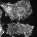
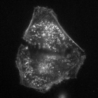
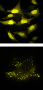
Visualizing the actin cytoskeleton in HeLa cells. The actin cytoskeleton is stained with rhodamine-phalloidin. Cells were either incubated with the a control (left) or the actin disrupting drug cytochalasin D (right). The control sample shows clear actin filaments (cables), whereas the drug-treated cells show a punctuate distribution of F-actin.
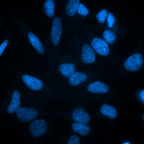
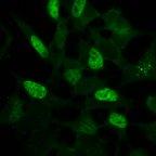
Detection of HeLa cells in the S-phase. Nuclei of all cells in the sample are stained with DAPI (blue fluorescent: left). S-phase cells are stained by incorporation and detection of BrdU (green fluorescence: right). For BrdU labeling, HeLa cells were incubated with BrdU for 15 minutes. Subsequently, immunofluorescence labeling is performed with a rat-anti-BrdU primary antibody and a goat-anti-rat-Alexa488 secondary antibody to detect BrdU. Note that some aspecific labeling of the cytoplasm is visible in the green channel.
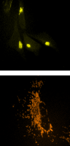
What do organelles look like in a eukaryotic cell? Several different plasmids encoding a fluorescent protein fused to an unkown organelle targeting signal are isolated and purified. The plasmids are transfected and the next day cells are observed with fluorescence microscopy. Students have to identify the organelle that is labeled with the fluorescent protein. Some examples of the organelle markers are shown here.